Nodule
A smaller focal opacity or lesion seen on imaging
Nodule is an imaging term for a smaller focal lesion or opacity seen on a chest image.
A nodule is a small focal spot or rounded opacity seen on imaging. Many are benign and stable, but some require follow-up depending on risk and appearance.
Representative X-ray
Illustrative reference image for this topic.
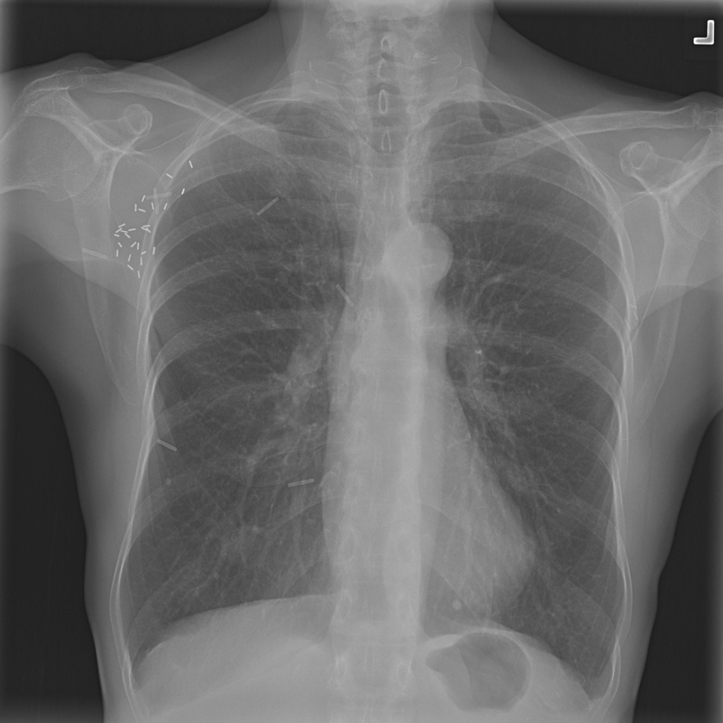
Reference image: PAT-894B · IMG-002 · Bounding-box highlight from source annotation where available.
What it is
- A nodule is a focal radiographic finding rather than a diagnosis
- It may reflect benign healed change, infection, inflammation, or malignant processes depending on size, morphology, and clinical context
How it appears on chest X-ray
- On chest X-ray, a nodule often appears as a relatively small rounded or oval opacity
- Visibility depends on size, contrast, location, and overlap with other structures
What radiologists look for
- Radiologists assess size, shape, margin, calcification pattern, location, and whether prior imaging or CT is needed to determine stability and significance
How X-ray helps
- Chest X-ray may be the first study to show a nodule, but CT is often needed for better characterization and follow-up planning
Common causes
- Possible causes include healed granulomas, infectious nodules, inflammatory change, benign tumors, or malignant lesions
Symptoms / associated symptoms
- Many nodules cause no symptoms
- Symptoms, when present, usually depend on the underlying cause rather than the nodule label itself
Risk factors
- Risk depends on the cause and can include smoking history, prior cancer, age, infection exposure, and environmental exposures
Why it can matter clinically
- A nodule can matter clinically because some need interval follow-up or additional imaging to determine whether they are stable or suspicious
When to seek medical care
- If a nodule is newly reported or associated with persistent symptoms, medical follow-up is appropriate
Evaluation and diagnosis
- Evaluation often includes prior-image comparison, CT, risk assessment, and follow-up planning based on the overall imaging and clinical picture
Treatment approaches
- Treatment depends on cause
- Some nodules simply need surveillance, while others need further diagnostic workup or treatment
Need help reviewing your own X-ray?
If you landed here because you are trying to understand a chest X-ray result, you can upload an image for an educational review and then use the related finding guides to go deeper.
FAQ
Is a lung nodule always dangerous?
No. Many nodules are benign, but some require follow-up depending on risk and imaging features.
Can chest X-ray fully characterize a nodule?
Often no. CT is commonly used for better detail.