Pneumothorax
Air in the pleural space that can partially or fully collapse a lung
Pneumothorax means air has collected in the pleural space and may partly or fully collapse the lung.
Pneumothorax means air is present around the lung instead of staying inside it. On X-ray, the key clue is often a visible pleural line with fewer lung markings beyond it.
Representative X-ray
Illustrative reference image for this topic.
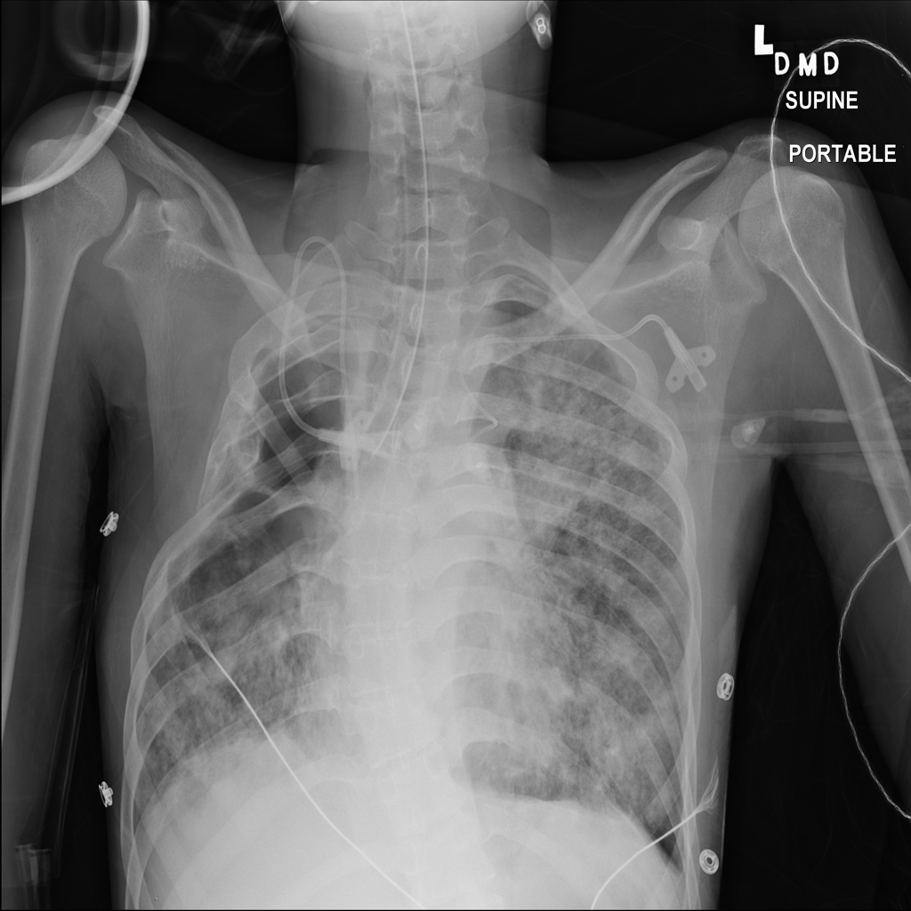
Reference image: PAT-EEB6 · IMG-029 · Bounding-box highlight from source annotation where available.
What it is
- Pneumothorax is air in the pleural space between the lung and chest wall
- On imaging, the key issue is separation between the lung edge and chest wall with reduced or absent normal lung markings beyond the pleural line
How it appears on chest X-ray
- On chest X-ray, pneumothorax may appear as pleural air with a visible visceral pleural line and relative absence of expected lung markings beyond that line
- Small pneumothoraces may be subtle, especially on portable or supine films
What radiologists look for
- Radiologists look for pleural air, the visible lung margin, degree of lung collapse, and whether there are signs of pressure effects such as mediastinal shift
How X-ray helps
- Chest X-ray can identify many pneumothoraces, estimate visible size, and help monitor interval change after treatment
- It can still miss small or position-dependent pleural air
Common causes
- Possible causes include spontaneous rupture of blebs, trauma, underlying lung disease, barotrauma, and medical procedures
- Some cases occur without an obvious trigger
Symptoms / associated symptoms
- Common symptoms can include sudden chest pain, shortness of breath, chest tightness, rapid breathing, and sometimes minimal symptoms if the pneumothorax is small
Risk factors
- Risk factors can include smoking, underlying lung disease, prior pneumothorax, trauma, positive-pressure ventilation, and recent invasive procedures involving the chest
Why it can matter clinically
- Complications can include worsening lung collapse, breathing difficulty, recurrence, and in more severe cases tension physiology with hemodynamic compromise
When to seek medical care
- Sudden chest pain, shortness of breath, worsening breathlessness, or concern for a new pneumothorax should be assessed promptly
- Severe breathing difficulty or instability needs urgent care
Evaluation and diagnosis
- Evaluation may include physical exam, chest X-ray, bedside ultrasound in some settings, oxygen assessment, and follow-up imaging depending on size and symptoms
Treatment approaches
- Management may include observation, oxygen support, needle aspiration, chest tube drainage, and follow-up imaging
- The right approach depends on size, symptoms, stability, and cause
Medication classes clinicians may use
Medications are usually supportive rather than curative for the air leak itself. Pain medicines and supportive medications may be used depending on symptoms and procedures performed.
Treatment modalities commonly paired with medication decisions
- observation
- oxygen support
- needle aspiration
- chest tube drainage
- specialist follow-up for recurrent cases
Pain relievers
Used to control chest discomfort and procedure-related pain.
- acetaminophen
- ibuprofen
- opioid analgesics in selected settings
Need help reviewing your own X-ray?
If you landed here because you are trying to understand a chest X-ray result, you can upload an image for an educational review and then use the related finding guides to go deeper.
FAQ
Can pneumothorax be missed on an X-ray?
Yes. Small pneumothoraces can be subtle, and technique or positioning can affect visibility.
Does a similar image mean I have a collapsed lung?
No. Similar-looking reference images do not diagnose your upload.